Goal
Regenerate a anatomically correct tooth directly in the patient's mouth using the patient's own stem cells
Problem
Loss of natural teeth and the limitations of conventional dental implants (long healing time, poor integration, cost)
Concept Summary
A three-dimensional, natural-material scaffold is loaded with growth factors (e.g., Wnt3a, BMP-7, VEGF, bFGF, NGF) and implanted into an empty tooth socket. The scaffold directs the patient's dental stem cells to migrate, proliferate, and differentiate into odontoblasts, dentin, cementum, and periodontal ligament, producing a functional tooth within weeks.
Detailed Description
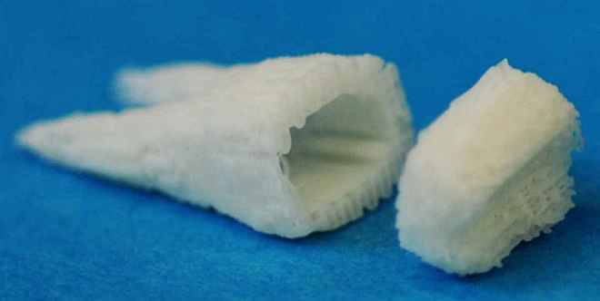
The technique, developed in Columbia University's Tissue Engineering and Regenerative Medicine Laboratory, uses an acellular, tooth-shaped scaffold made from biocompatible natural materials. The scaffold is pre-infused with a cocktail of signaling proteins that promote odontoblastic differentiation and tissue mineralization. After surgical placement into the extraction socket, endogenous stem cells from the dental pulp or surrounding tissues colonize the scaffold, forming dentin, enamel, cementum, periodontal ligament, and alveolar bone. Animal studies have shown formation of anatomically correct tooth-like structures in vivo within approximately nine weeks. Patents have been filed covering the scaffold composition, growth-factor combinations, and methods of use.
Principles
- Stem-cell guided tissue engineering
- Growth-factor mediated cellular differentiation
- Three-dimensional scaffold architecture for spatial control
Scientific Domains
Materials
- Natural biocompatible scaffold material (e.g., collagen, hydrogel, decellularized tooth matrix)
- Growth factors: Wnt3a, BMP-7, VEGF, bFGF, NGF
- Acellular tooth-shaped scaffold
Mechanisms of Action
- Scaffold provides a 3-D template for cell attachment and organization
- Wnt3a and BMP-7 induce odontoblastic lineage commitment
- VEGF, bFGF, NGF support angiogenesis, cell migration, and nerve integration
- Endogenous dental stem cells populate the scaffold and produce mineralized dental tissues
Applications
- Clinical tooth regeneration for edentulous patients
- Periodontal tissue repair
- Alveolar bone regeneration
Claimed Performance
Anatomically correct tooth-like structures formed in vivo within nine weeks after implantation in animal models.
Experimental Evidence
Animal-model studies reported successful in-situ regeneration of tooth-like structures, periodontal ligament, and alveolar bone using the scaffold-stem-cell approach; multiple patents have been filed describing the method.
Replication Status
Demonstrated in animal models; not yet validated in human clinical trials.
Limitations
- Not yet approved for human use; regulatory hurdles remain
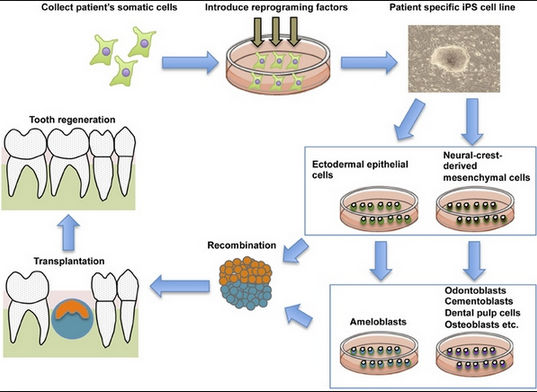
- Requires harvesting and processing of patient-specific stem cells
- Potential high cost of scaffold manufacturing and growth-factor cocktails
- Long-term durability and functional integration of regenerated teeth are unproven