Goal
Photograph sub-atomic and molecular structures in true color with high resolution and penetration.
Problem
Electron microscopes provide only black-and-white images, limited penetration of internal structure, and require complex preparation.
Concept Summary
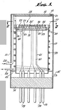
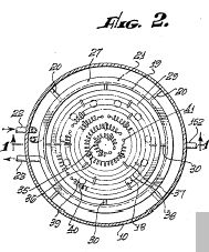
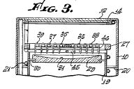
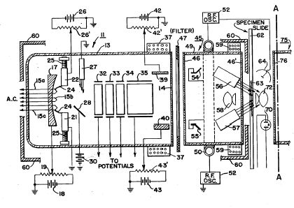
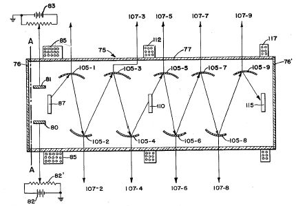
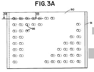
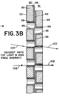
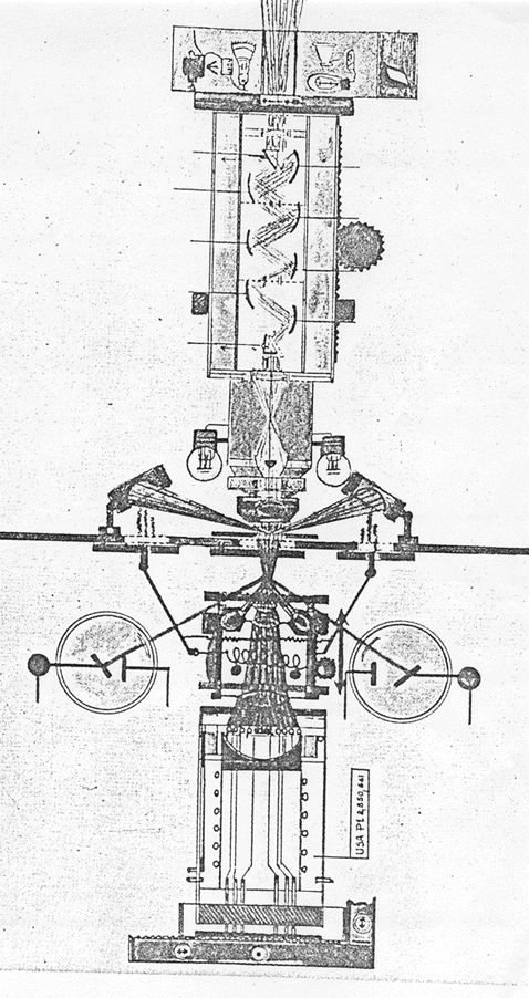
The Nemescope uses multiple radiation sources (cold-cathode lamp, radium guns, high-frequency coils) to bombard a specimen, causing it to emit resonant frequencies that are converted to visible light in an orthicon tube, producing high-magnification color images.
Principles
- Radiation excitation of specimens
- Multiple radiation sources with slightly different frequencies
- Resonant frequency emission
- Optical conversion and amplification via orthicon tube
Scientific Domains
Materials
- lead
- platinum
- gold
- germanium
- tungsten
- radium
- quartz
- semiprecious stones
- steel
Mechanisms of Action
- Bombardment of specimen with radiation (electron, alpha, beta, gamma)
- Emission of ultra-spectral light at resonant frequencies
- Conversion of emitted radiation to visible light
- Optical magnification and projection
Energy Sources
Applications
- Medical diagnostics
- Materials analysis
- Metallurgy
- Virology
Claimed Performance
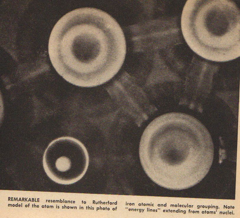
Resolution down to atomic nucleus, projected magnification up to 5 million X, true-color imaging of cells, enzymes, metals, and viruses.
Experimental Evidence
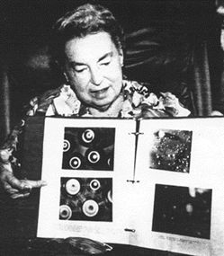
Images of blood and urine cells from cancer patients, resolved enzymes, metallic alloy fault lines, atomic nucleus structures, and virus particles were reported.
Limitations
- Requires complex and potentially hazardous radiation sources
- No independent verification or peer-reviewed data
- Safety concerns from high-energy radiation
Red Flags
- Extraordinary claims without quantitative data
- Device was reportedly stolen and not publicly demonstrated
- Potential safety hazards from radiation exposure